A new Society of Radiological Ultrasound (SRU) expert consensus statement on the topic of screening for endometriosis has been published in the journal Radiology. Various procedures are recommended for symptomatic patients or those at high risk of endometriosis.
Endometriosis is a disease with significant delays in diagnosis and medical imaging, if done uniformly and up to date, can benefit patients suffering from it.
There is a seven-year delay between the onset of symptoms and diagnosis of endometriosis.
That’s why the Society of Radiological Ultrasound (SRU) has just publish a consensus statement in the journal Radiology its experts to disseminate best practice recommendations. Endometriosis, characterized by the presence of endometrium-like tissue outside the uterus, affects 10% of women of reproductive age and occurs in 21% of women who have had a hysterectomy accompanied by chronic pelvic pain. The time between the onset of symptoms and diagnosis of endometriosis is estimated to be seven years.
Endometriosis is also associated with infertility and subfertility, affecting 20-50% of patients with these conditions. Ultrasound is typically the first-line imaging modality used when patients report chronic pelvic pain or have infertility problems, but it appears to be underutilized for screening for deep endometriosis.
An expert panel is set to publish recommendations on best imaging practices for diagnosing endometriosis.
![]() The SRU therefore convened a multidisciplinary panel of experts to provide recommendations to improve the endometriosis screening process. “The goal of this consensus group is to recommend methods that increase the diagnostic sensitivity of endometriosis to pelvic ultrasound by increasing awareness, improving interpretation, adding simple, high-throughput methods for determining the depth of endometriosis, and improving triage protocols,” says the study’s first author. document, Dr. Scott W. Young, consultant in diagnostic radiology, department of ultrasound, Mayo Clinic in Phoenix (Arizona, USA).
The SRU therefore convened a multidisciplinary panel of experts to provide recommendations to improve the endometriosis screening process. “The goal of this consensus group is to recommend methods that increase the diagnostic sensitivity of endometriosis to pelvic ultrasound by increasing awareness, improving interpretation, adding simple, high-throughput methods for determining the depth of endometriosis, and improving triage protocols,” says the study’s first author. document, Dr. Scott W. Young, consultant in diagnostic radiology, department of ultrasound, Mayo Clinic in Phoenix (Arizona, USA).
The panel consisted of experts in the imaging and treatment of endometriosis, including radiologists, sonographers, gynecologists, reproductive endocrinologists, and minimally invasive gynecologic surgeons. A comprehensive literature review combined with a modified Delphi technique resulted in consensus.
“The statement identifies the target population for screening, describes modalities complementary to pelvic ultrasound, establishes direct and indirect ultrasound surveillance of endometriosis, establishes a system for evaluating and reporting observations, and makes recommendations for additional imaging and specific patient care.” , continues Dr. Young.
Recommendations for additional imaging depending on the type of patient
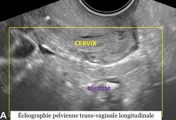
The panel’s recommendations include transvaginal posterior ultrasound, observation of the relative position of the uterus and ovaries, and modification of the uterine sliding sign to improve detection of endometriosis. “These additional techniques are typically performed in less than five minutes and may ultimately reduce the time to diagnose endometriosis in at-risk patients,” adds Dr. Young.
![]() The committee also recommends that direct and indirect evidence of deep endometriosis be assessed during the examination and results reported in four categories: incomplete (APU-0), normal (APU-1), equivocal (APU-2), and positive (APU-2). -3), with appropriate management recommendations.
The committee also recommends that direct and indirect evidence of deep endometriosis be assessed during the examination and results reported in four categories: incomplete (APU-0), normal (APU-1), equivocal (APU-2), and positive (APU-2). -3), with appropriate management recommendations.
Focus on deep endometriosis research
“The SRU consensus on routine pelvic ultrasound for endometriosis aims to improve detection of deep endometriosis even during initial ultrasound, with minimal additional time during imaging and without special patient preparation,” concludes Dr Young. Focusing imaging on anatomical areas where deep endometriosis is common may improve detection and reduce diagnostic time. »
These recommendations are intended for symptomatic patients at typical risk for endometriosis. Patients at high risk due to previous diagnostic or therapeutic laparoscopy for endometriosis or serious clinical indications may directly benefit from advanced imaging of endometriosis, especially if they are likely to undergo surgery or if monitoring is necessary in the context of infertility and medical treatment.
The authors point out that validation studies will be needed to prove the accuracy of these recommendations and to ensure their routine implementation.
Bruno Behnke with RSNA
Related Posts

PROSTATE CANCER: Plant foods against its progression
Analyze your breathing to monitor your health in intensive care