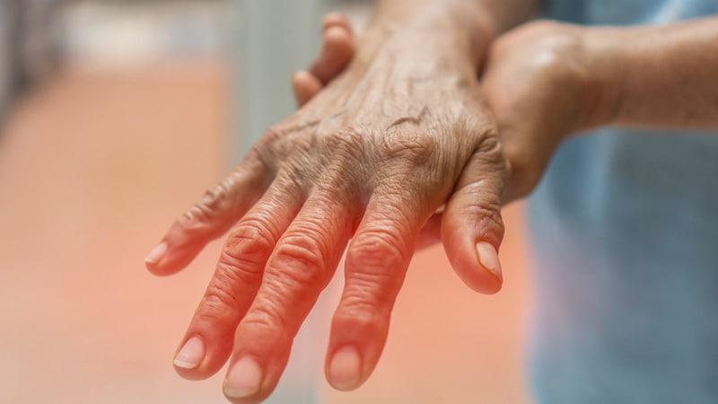
Toulouse, France – Diabetic neuropathy was the topic of a plenary session dedicated to the congress of the Francophone Society of Diabetes.[1]. The reason is simple: this already very common problem is progressing and remains insufficiently addressed. Phenotyping patients is still necessary, and for this purpose the use of neurofilaments does not solve everything, and EMG can be misleading, the researcher explains. Previously Agnes HartemannHead of the Department of Diabetology at the Pitié-Salpêtrière Hospital (APHP, Paris).
“The number of people with diabetic neuropathy worldwide has more than tripled since 1990, reaching 206 million in 2021,” it said. Pre Liana Ongfrom the University of Washington (Seattle, USA) and co-author of the study Global Burden of Disease, Injury and Risk Factors (GBD) Study in 2021[2].
Neuropathic pain, according to the literature, appears in 25–30% of people with diabetic peripheral neuropathy. [3]. Not surprisingly, this progresses with age: over the 26 years of study follow-up Epidemiology of Diabetes Interventions and Complications (EDIC)observational monitoring study DCCT (1982–1993), prevalence of neuropathic pain (Q2: “ Have you ever felt a burning sensation in your legs and/or feet? ? ” and/or question 6: ” Does it hurt when the blanket touches your skin?“) increased from 8.5% to 19.8%, while the Michigan Neuropathy Screening Instrument/MNSI score greater than 2 increased from 22.9% to 43.5%. [4].
Diabetic neuropathy can no longer be equated with small fibers.
According to Professor Agnes Hartemann, who presented the pathophysiology of neuropathic pain (NP), “monofilaments have taken an excessive place in screening.” She explains: “It has long been thought that sensory neuropathy affects small fibers and painful neuropathy affects large fibers. However, this distinction no longer holds because in both types of neuropathy, both types of fibers can be damaged. “In peripheral neuropathy, there are actually two forms of nerve damage: on the one hand, loss of fibers leading to loss of function (so-called “sensory” neuropathy), and, on the other hand, neuropathy with overactive fibers. , hyperexcitability, which represents an increase in function. This involves dysfunctional ion channels with spontaneous, iterative, untimely activation at the peripheral level with consequences at spinal cord connections.
What are the signs and symptoms of loss and gain of function?
Loss of function (sensory neuropathy) corresponds to a rarefaction of nerve fibers, both large (>30 m/s) and small (3-30 m/s for weakly myelinated and <3 m/s for non-myelinated). “When you look for loss of large fiber function, you will find loss of osteotendinous reflexes, decreased vibration perception and proprioception, sensitivity to touch and pressure,” she explains. This is what only 10g monofilament probes: light touch, between touch and pressure. “For its part, the rarefaction of small fibers leads to a decrease in sensitivity to pain (needle test), the perception of hot and cold, as well as sensitivity to pressure; this appears to be common for large and small fibers.
Moreover, painful neuropathy (loss of function) does not only affect small fibers, as previously thought, since hyperexcitability “can originate from large fibers,” points out Agnès Hartemann. Thus, patients describe a feeling as if the foot is being squeezed in a vice, as well as mechanical allodynia (rubbing with sheets or cotton wool). As for increased excitability at the level of small fibers, it causes the well-known symptoms of stinging, painful cold (the sensation of walking barefoot in the snow), burning, itching, thermal allodynia, hyperalgesia, as well as electrical discharges, the latter predominantly arising from small fibers.
There is also a loss of pain inhibitory function at the level of the spinal cord. Increased arousal affects the brain, leading to increased depression, anxiety, and sleep disturbances secondary to pain. However, the frequency and duration of these disorders exceed what is observed with chronic pain of the same intensity, but of a different origin, with amplification due to the vicious peripheral/medullary/central circle.
As for whether neuropathy begins with fiber overactivity or loss of function, it is not so clear. The percentage of patients with one, the other, or both neuropathies depends on the population studied and the instruments used. In one study among others that included 232 patients with type 1 and type 2 diabetes (74%) with a mean age of 63 years and neuropathy confirmed by electromyography (EMG) or biopsy, researchers found deafferentation in 54% of “irritable nociceptors” ” in 15% and both in 31% [5].
EMG, only in case of doubt about the diagnosis
The diagnosis of rarefied fibers, that is, sensory neuropathy, is essentially clinical. The electromyogram may be abnormal only if the loss of function affects large fibers. Therefore, without EMG abnormalities, one may erroneously conclude that diabetic neuropathy is not present, even in the presence of targeted small fiber damage.
Skin biopsy of the ankle, which reveals thinning of small fibers of the epidermis and dermis, continues to be used in clinical trials to determine the phenotyping of patients. As for confocal microscopy of the cornea (indirect vision of the loss of small fibers), it has not yet been standardized to date.
In turn, the diagnosis of hyperactivity (excitability) is also clinical. EMG, skin biopsy, and corneal confocal microscopy may be normal and therefore unnecessary for a positive diagnosis. “We must send our patients to CETD pain centers so that they are phenotyped and receive the treatment most appropriate for the type of pain,” the diabetologist insists. Recognizing DN is critical, especially in patients with diabetes, who may suffer from a variety of pain, especially in the lower extremities. »
In this regard, screening questionnaire DN4 [6] has been reconfirmed specifically and by several diabetic neuropathy groups. [7]. Grade > 4 causes DN with a sensitivity of 83% and specificity of 90%.
A study published in 2013, in which Agnes Hartmann participated, found 14% of patients with type 1 diabetes and 24% of patients with type 2 diabetes with DN. 70% sought advice for pain, but only 38% received appropriate treatment.
Certain characteristics may cast doubt on the diagnosis of diabetic neuropathy, especially rapid onset, symmetry, severe motor deficits, or proximal involvement, which warrant referral to a neurologist.
As for complex diagnoses, these may include radiculopathies associated with the cervical, spinal and lumbar regions. In these cases, EMG and MRI are relevant. Other etiologic factors to consider include post-stroke neuropathy, Parkinson’s disease, chemotherapy, even knee osteoarthritis or lower extremity occlusive arteriopathy.
Where do loss of function and hyperactivation neuropathies come from?
Neuropathy is characterized by microangiopathy resulting from damage to the microvessels innervating the nerves. But neuropathy has many risk factors, including blood sugar and metabolic syndrome. [8], but also overweight, cardiovascular disease, dyslipidemia, high blood pressure and smoking. “It even starts with prediabetes type 2,” adds Professor Hartemann. Thus, there is an influence of chronic hyperglycemia (microangiopathy of the endonervous capillaries) as well as axonal insulin resistance associated with the same risk factors as for muscles. We see mitochondrial axonal dysfunction, oxidative stress and reticulum stress.”[9].
Links of interest from experts:
– Agnes Hartemann: No
Register for Medscape Newsletters : select your choice


